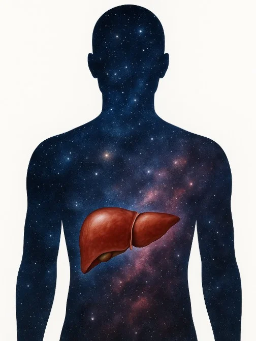
Introduction: The Silent Epidemic
In the vast universe of human health our liver often operates silently and diligently, performing over 500 vital functions. In recent decades, a stealthy adversary has emerged: Non-Alcoholic Fatty Liver Disease (NAFLD). NAFLD is affecting approximately 25% of the global population, and it is directly linked to the modern lifestyle: sedentary habits, calorie-dense diets, and the metabolic syndrome constellation. [1]
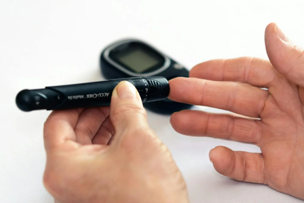
As opposed to liver diseases caused by alcohol or viruses, NAFLD creeps in without overt symptoms, making early detection very challenging. But luckily, our body’s biochemistry offers some clues, most notably through liver enzymes like Alanine Aminotransferase (ALT) and Aspartate Aminotransferase (AST).
Meet the Enzymes: ALT and AST
ALT and AST are enzymes mainly found in liver cells and they play crucial roles in amino acid metabolism. “How they end up in our blood?” would probably be your next question. Well, the answer is note that complicated – When liver cells are damaged or inflamed, these enzymes leak into the bloodstream, elevating their serum levels.
- ALT (Alanine Aminotransferase): Primarily located in the liver, making it a more specific indicator of liver injury.
- AST (Aspartate Aminotransferase): Found in the liver, heart, muscles, kidneys, and brain, thus less specific to the liver.
Elevated levels of these enzymes, a condition termed “transaminitis,” signal liver distress but don’t pinpoint the exact cause.
There’s a ‘dad joke’ about ALT and AST, which might help you remember what these two enzymes are doing:
AST: “Hey ALT, you only care about the liver!”
ALT: “And you? You’re just all over the place!”
NAFLD: A Modern Metabolic Challenge
NAFLD is a spectrum of disorders: from simple steatosis (fat accumulation in the liver) to Non-Alcoholic Steatohepatitis (NASH), which involves inflammation and can progress to fibrosis, cirrhosis, or even liver cancer.
Risk Factors:
- Obesity: Excess fat, especially visceral fat, contributes to liver fat accumulation.
- Type 2 Diabetes: Insulin resistance promotes fat deposition in the liver.
- Dyslipidemia: High triglycerides and low HDL cholesterol levels are common in NAFLD patients.
- Metabolic Syndrome: A cluster of conditions—including hypertension, high blood sugar, excess body fat around the waist, and abnormal cholesterol levels—increases NAFLD risk.
 Interestingly, many individuals with NAFLD remain asymptomatic, and the disease is often discovered incidentally during routine blood tests or imaging studies.
Interpreting Enzyme Levels in NAFLD
In NAFLD, ALT levels are typically higher than AST levels, with an AST/ALT ratio less than 1. This contrasts with alcoholic liver disease, where AST often exceeds ALT, and the ratio is greater than 2.
However, it’s crucial to note that normal ALT and AST levels don’t necessarily rule out NAFLD. Studies have shown that individuals can have significant liver fat accumulation and even inflammation despite normal enzyme levels. [2]
Therefore, while elevated transaminases can hint at liver issues, they are not definitive diagnostic tools for NAFLD.
Beyond ALT and AST: Additional Diagnostic Tools
Given the limitations of ALT and AST, clinicians employ various tools to assess liver health:
- Imaging Studies:
- Ultrasound: Often the first imaging modality used; can detect liver steatosis when fat content exceeds 20-30%.
- MRI and CT Scans: Provide more detailed images and can quantify liver fat content.
- Non-Invasive Scoring Systems:
- FIB-4 Index: Calculates fibrosis risk based on age, AST, ALT, and platelet count.
- NAFLD Fibrosis Score: Incorporates multiple variables to estimate fibrosis stage.
- Liver Biopsy: Considered the gold standard for diagnosing NASH and assessing fibrosis but is invasive and not routinely performed.
 Management and Treatment Strategies
 Management and Treatment Strategies
Addressing NAFLD involves a multifaceted approach:
- Lifestyle Modifications:
- Weight Loss: A reduction of 7–10% in body weight can significantly improve liver histology. [3]
- Diet: Adopting a Mediterranean diet rich in fruits, vegetables, whole grains, and healthy fats.
- Exercise: Regular physical activity enhances insulin sensitivity and reduces liver fat.
- Pharmacological Interventions:
- Vitamin E: Antioxidant properties may benefit non-diabetic patients with NASH. [4]
- Pioglitazone: A diabetes medication that has shown promise in improving liver histology in NASH patients.
- GLP-1 Receptor Agonists: Originally for diabetes, these drugs aid in weight loss and may benefit liver health. [5]
It’s essential to tailor treatment plans to individual patient profiles, considering comorbidities and potential drug side effects.
Conclusion: Navigating Liver Health
The liver, our body’s metabolic powerhouse, often suffers silently. NAFLD, a manifestation of modern lifestyle challenges, underscores the importance of proactive health monitoring. While ALT and AST provide valuable insights, they are pieces of a larger puzzle. Comprehensive evaluation, early detection, and holistic management are paramount.
As we continue to explore the intricacies of liver health, embracing a proactive approach can ensure our livers remain resilient in the face of modern challenges.
References
- Younossi ZM et al. (2016). Global epidemiology of NAFLD. Hepatology.
- Tapper EB, Lok AS. (2018). Misconception: You Can’t Have Liver Disease With Normal Liver Tests. Clinical Liver Disease.
- Vilar-Gomez E et al. (2015). Weight loss improves NASH. Gastroenterology.
- Sanyal AJ et al. (2010). Vitamin E for NASH. NEJM.
- Newsome PN et al. (2021). Semaglutide in NASH. The Lancet.